
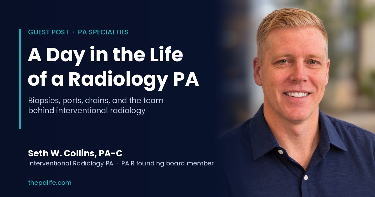
This guest post was written by Seth W. Collins, PA-C, an interventional radiology PA and founding board member of Physician Associates in Radiology (PAIR).
When most people think about physician associates, they picture primary care clinics, emergency departments, or operating rooms. Radiology tends to exist somewhere in the medical subconscious, vaguely understood as the darkened room where someone squints at CT scans while drinking coffee of questionable quality.
And to be fair, some of that is true.
But modern radiology, particularly interventional radiology, is also filled with procedures, patient care, imaging technology, anatomy, pathology, critical thinking, and just enough controlled chaos to keep things interesting.
7:30 AM: Reviewing the Day's Schedule
I work as a physician associate in interventional radiology (IR), and my day usually starts around 7:30 in the morning with a review of the procedural schedule. The list is already impressively eclectic before the coffee has fully taken effect.
There are outpatient biopsies, vascular access procedures, fluoroscopic studies, drainages, injections, and a collection of inpatient add-ons that materialized overnight. These inpatient consults are often patients admitted to the hospital whose teams have identified a possible need for an IR procedure. Part of my job is reviewing those requests to determine whether the procedure is appropriate, indicated, and safe to perform.
Sometimes the answer is straightforward. Sometimes it requires a surprising amount of detective work involving imaging, lab values, medical history, anticoagulation status, and deciphering consult notes written at 2 AM. If the procedure is appropriate, I coordinate with our nursing and scheduling teams to get the patient added to the schedule, often the very same day.
Before heading down to see my first patient, I stop by the interventional radiologist's office to say good morning. He is already reviewing the first few cases of the day, and we briefly look over the imaging for a patient he is about to treat. He is performing a kyphoplasty, a minimally invasive procedure used to stabilize a painful vertebral compression fracture. The goal is to guide a needle into the collapsed vertebra, inflate a small balloon to make a little room, and then fill the resulting cavity with bone cement to stabilize the fracture. We talk through the case for a minute, point out a few things on the imaging, and then go our separate ways to start the morning.
First Case: A CT-Guided Lung Biopsy
My first case is a CT-guided lung biopsy on a 72-year-old gentleman with a greater than 40-pack-year smoking history. A recent CT scan demonstrated a 2.2-centimeter spiculated nodule in the right lower lobe of his lung. "Spiculated," for those mercifully not yet buried in medical terminology, essentially means the nodule isn't content to sit there with a nice, clean border. It's pulling and distorting the surrounding lung, creating those characteristic radiating strands. It's not proof of cancer, but it is one of those CT findings that makes us considerably more concerned.
Before the procedure, I meet the patient in the pre-procedure area. One of the misconceptions about radiology is that it lacks patient interaction. In reality, much of my day involves meeting patients during vulnerable and often frightening moments in their lives.
I review his medical history, perform a physical exam, assess his airway, and assign an ASA score, which is a classification system used to estimate procedural risk related to anesthesia and sedation. We then discuss informed consent, which in medicine means far more than obtaining a signature on a form.
A proper informed consent discussion involves explaining the purpose of the procedure, expected benefits, alternatives, and possible complications in a way that allows the patient to make an educated decision. Medicine is filled with astonishing technology, but occasionally the most important thing you can do is simply sit down and explain things clearly to another human being.
Once consent is completed, the patient is transferred to the CT scanner and positioned prone, meaning on his stomach. Preliminary CT images are obtained to localize the lesion and compare it to prior imaging. Occasionally abnormalities improve or resolve entirely, sparing patients from invasive procedures altogether. Unfortunately, the nodule remains conspicuously present and continues to make a persuasive case for biopsy.
Before beginning, we perform a timeout, a universal patient safety pause where the entire team confirms the patient's identity, allergies, imaging, anticoagulation status, and the planned procedure. It is one of the many things modern medicine does exceedingly well, recognizing that humans, while generally competent, are also capable of spectacular errors when left unchecked.
Inside the CT procedure room are the patient, myself, and a registered nurse providing medications and monitoring. Behind the glass in the control room is the CT technologist, operating the scanner and helping coordinate imaging and supplies.
The IR procedure rooms function much like operating rooms: the nurse and I wear lead protective gear, and I scrub in before putting on a sterile gown and gloves. Once the patient is adequately sedated with fentanyl and Versed, I sterilize the skin, place sterile drapes, numb the tissues with local anesthetic, and begin advancing a biopsy needle toward the lung lesion using CT fluoroscopy for real-time imaging guidance.
There is something uniquely satisfying about image-guided procedures. The work involves navigating anatomy, reading imaging on the fly, and advancing instruments with millimeter precision, while trying to preserve the useful distinction between access and damage.
Using a spring-loaded biopsy device, I obtain several core tissue samples. At our institution, a pathologist comes directly into the procedure suite to evaluate the specimens in real time. The goal is not necessarily to establish a diagnosis immediately, but rather to confirm that adequate tissue has been obtained and processed correctly.
Once the samples are collected, I remove the needle and obtain additional CT imaging to evaluate for complications. One of the most common risks after lung biopsy is a pneumothorax, which occurs when air leaks from the lung into the pleural space surrounding it. Small pneumothoraces may simply require observation, while larger ones occasionally require placement of a chest tube.
Fortunately, today's post-procedure imaging demonstrates only mild expected post-biopsy changes and a small amount of localized bleeding within the lung tissue. The patient remains stable and comfortable and is transferred to recovery.
As he heads to the recovery unit, I head back to my office to enter post-procedure orders, including monitoring parameters, sedation documentation, and a follow-up chest X-ray to ensure no delayed pneumothorax develops prior to discharge.
Mid-Morning: Port Placement and an Esophagram
By this point, my next patient has already arrived. She is a middle-aged woman with metastatic colon cancer referred for placement of an implanted venous port prior to chemotherapy.
A port is a small device implanted beneath the skin, usually in the upper chest, connected to a catheter that terminates in a large central vein near the heart. These devices provide durable long-term vascular access for chemotherapy, immunotherapy, lab draws, and other treatments. They are also vastly preferable to repeatedly poking someone's arm veins every few days for months on end, which patients tend not to enjoy.
An IR suite is an impressive collection of machinery whose principal purpose is to allow us to look inside people without opening them up, which is a considerable improvement over the old-fashioned approach. We use ultrasound for real-time imaging and a fluoroscopy unit for live X-ray images. The fluoroscopy machine is mounted on a large C-shaped arm that can swing around the patient and be positioned at different angles, allowing us to examine the same anatomy from several directions. The IR technologist and I operate the equipment from within the room, moving the machine, adjusting the imaging field, and obtaining the views we need as the procedure progresses. It is an extraordinary piece of technology, and one that has made it possible to perform increasingly complex procedures through remarkably small openings.
Using ultrasound guidance, I access the internal jugular vein in the neck. Fluoroscopy, essentially live X-ray imaging, allows me to guide the catheter into precise position where the superior vena cava joins the right atrium. The procedure itself takes roughly 20 minutes, after which the catheter position is confirmed, the small incisions are sutured and dressed, and the patient is transferred to recovery.
Later that morning, I transition briefly into diagnostic radiology for an esophagram study.
The procedure uses fluoroscopic imaging to observe the esophagus as the patient drinks contrast material, revealing that even the simple act of swallowing contains a remarkable amount of choreography.
This particular patient has longstanding refractory heartburn symptoms despite medical therapy. As she swallows contrast, we observe a moderate to large paraesophageal hernia, where part of the stomach protrudes upward through the diaphragm into the chest cavity.
Late Morning: A Renal Biopsy
From there, I head over to ultrasound for a non-targeted renal biopsy on a patient with worsening kidney function of unclear etiology.
Unlike a targeted biopsy where we sample a visible lesion, this procedure involves obtaining tissue from the lower pole renal cortex, the outer portion of the kidney containing the glomeruli, the microscopic filtration units responsible for cleaning the blood.
After obtaining informed consent and initiating moderate sedation, I use real-time ultrasound guidance to advance the biopsy needle into the lower pole cortex while carefully avoiding major vessels and the central collecting system.
The samples will ultimately be sent to a specialized renal pathology center for advanced analysis, helping determine the precise mechanism of the patient's kidney disease.
Lunch: The Team Behind the Procedures
By the time I finish documentation and post-procedure orders, it's a little past noon.
Having once again failed to pack a lunch, a shortcoming that seems to resist correction by either experience or intent, I make my way to the cafeteria. I meet up with two of our IR technologists, the same ones who assisted with the port placement earlier that morning, and we grab a quick lunch.
One of the underrated aspects of radiology is how collaborative it is. Good procedural days depend heavily on strong relationships between physicians, PAs, nurses, technologists, recovery staff, and schedulers. It is the kind of work where efficiency is rarely the product of any single person, but rather of a shared rhythm that develops over time, where people anticipate each other's needs, compensate for small gaps, and keep the entire process moving without anyone needing to announce what they are doing.
As we finish eating, the lead technologist gets a text letting us know our next patient is running a few minutes late. We briefly take advantage of the unexpected downtime and go for a quick walk around the hospital campus before the afternoon cases begin.
Afternoon: Epidural Steroid Injection
Not long afterward, the patient arrives for the epidural steroid injection.
The patient had developed persistent lower back pain with radicular symptoms, meaning pain radiating down the leg due to nerve irritation. MRI imaging demonstrated degenerative disc disease, and several weeks of physical therapy had failed to improve symptoms.
Under fluoroscopic guidance, I advance a thin spinal needle between the L4 and L5 vertebral bodies into the epidural space, carefully navigating between bony landmarks and soft tissue. The needle is positioned just outside the dural sac, avoiding the cerebrospinal fluid, in order to allow targeted delivery of medication around the affected nerve roots.
Once needle position is confirmed using a small amount of contrast in what is called an epidurogram, I inject a combination of anesthetic and corticosteroid. The anesthetic provides more immediate temporary relief, while the steroid works gradually over several days to reduce inflammation surrounding the affected nerves.
The ultimate goal is not simply pain reduction, but helping the patient regain enough function to participate more effectively in physical therapy and return to normal daily life.
An Inpatient Add-On: Hepatic Abscess Drainage
After finishing documentation, I ask the radiologist to take the next outpatient case so I can move on to an inpatient add-on. He graciously agrees, and appears quietly pleased at the chance to step away from the increasingly robust stack of diagnostic studies waiting in the queue.
My next case is a hepatic abscess drainage in a patient who presented with fever, abdominal pain, and an elevated white blood cell count. CT imaging demonstrated a characteristic rim-enhancing fluid collection within the liver, often the hallmark appearance of an abscess, sometimes accompanied by surrounding edema or internal complexity that helps distinguish it from other hepatic lesions. In this setting, the diagnosis of a liver abscess was most likely.
Using CT guidance, I advance a small needle through the liver and into the abscess cavity. Once access is established, I pass a guidewire through the needle and coil it within the collection. Removing the needle leaves the wire in place, allowing me to pass dilators and ultimately place a drainage catheter directly into the abscess.
Almost immediately, purulent fluid begins draining from the catheter.
I collect samples for microbiology culture so the infectious disease team can tailor antibiotic therapy appropriately. Assuming the patient improves clinically and follow-up imaging demonstrates resolution, I will likely see him again in a few weeks to evaluate and potentially remove the drain.
Shortly after the procedure, the patient develops transient hypotension and rigors, severe shaking chills that can occur when manipulating an abscess and temporarily releasing bacteria into the bloodstream. We administer IV fluids and medications while closely monitoring him. Fortunately, he stabilizes appropriately and avoids requiring transfer to a higher level of care.
Late Afternoon: Genicular Nerve Ablation and Arthrograms
The next patient is known to me from prior clinic visits and procedures. She is scheduled today for a genicular nerve ablation. She has previously undergone a total knee replacement but continues to experience significant chronic knee pain despite having exhausted several other treatment options.
Genicular nerve ablation is a minimally invasive procedure designed to interrupt some of the sensory nerves responsible for transmitting pain from the knee. Using fluoroscopic guidance, I identify the appropriate bony landmarks around the knee and place probes near the target nerves. After confirming their positions, the nerves are treated with radiofrequency energy, creating a controlled lesion that disrupts their ability to transmit pain signals.
It is a relatively small procedure for a problem that can have an enormous effect on someone's daily life. The goal is not to rebuild the knee or eliminate every sensation of discomfort, but to reduce pain enough that the patient can walk more comfortably, sleep better, and return to activities that chronic pain has gradually pushed out of her life.
By late afternoon, I finish the procedural day with several pre-MRI arthrogram injections.
These are relatively quick fluoroscopic procedures where contrast is injected directly into joints, commonly the shoulder or hip, prior to MRI imaging. By introducing diluted gadolinium into the joint itself, the subsequent MRI can provide exquisite detail when evaluating structures such as the labrum, cartilage, ligaments, and rotator cuff tendons.
End of the Day: Reports and Protocols
As the schedule finally winds down, I return to my office to dictate reports for the day's studies and procedures. Documentation, while rarely glamorous, is a critically important part of radiology practice.
Before heading home, I review upcoming outpatient procedures for the next several days. This process, often called protocoling, involves reviewing imaging and clinical history to determine whether the procedure requested is the most appropriate for the patient.
As I finish signing protocols and handing cases off to the schedulers, the nurses and technologists stop by to say goodnight on their way out. I check in briefly with the radiologist, who is wrapping up his own work, and we all head out the door together.
And that is another day in radiology.
Why PAIR Exists
One of the things I have learned throughout my career is that no two radiology PA practices look exactly the same. Some PAs work primarily in interventional radiology, others in neurointerventional radiology or diagnostic imaging, and many cover a combination of roles. Some work in large academic centers with extensive PA teams, while others function as solo practitioners with few, if any, PA colleagues nearby.
That diversity is one of the strengths of the field, but it also creates real challenges when it comes to consistency, mentorship, and professional support. PAIR, Physician Associates in Radiology, was created to meet that need, providing a unified organization that connects PAs across all practice environments and strengthens the profession as a whole. Its mission is to build community, mentorship, advocacy, education, and a stronger professional identity for PAs working across radiology specialties.
As PA roles within radiology continue to expand, it has become increasingly apparent that there are limited spaces dedicated specifically to bringing radiology PAs together. PAIR was created to help fill that space and give PAs working in radiology an opportunity to learn from one another, build professional relationships, and have a collective voice within the specialty.
For students interested in anatomy, procedures, imaging, technology, critical thinking, and collaborative medicine, radiology offers an incredibly rewarding and dynamic career path that many people simply do not realize exists.
And tomorrow morning, when I walk back into the department, I already know the day will look completely different all over again.
About the Author

Seth W. Collins, PA-C, is a physician associate specializing in interventional radiology and serves as Manager of the Interventional Radiology Advanced Practitioner Service Line at Jefferson Radiology. He leads the development and integration of physician associates within radiology, with a focus on education, clinical practice, and expanding advanced practice roles in interventional radiology.
He is a founding board member of Physician Associates in Radiology (PAIR), where he works to advance the profession through education, advocacy, and professional development. He earned his Master of Science in Physician Assistant Studies and Bachelor of Science in Health Studies from Springfield College.
You may also like -
 Overcoming Obstacles on Your Path to Becoming a Physician Assistant
Life is full of obstacles. They can be as simple as getting out of bed in the morning or as arduous as writing the perfect PA school application essay.
Most of the time, the biggest obstacle is ourselves (affiliate link). The […]
Overcoming Obstacles on Your Path to Becoming a Physician Assistant
Life is full of obstacles. They can be as simple as getting out of bed in the morning or as arduous as writing the perfect PA school application essay.
Most of the time, the biggest obstacle is ourselves (affiliate link). The […]- Is Physician Assistant (PA) School Hard?
Is PA school hard?
Here is a question I get asked all the time: Is PA school difficult?
Of course, in many ways, it is equally as rigorous as medical school.
The didactic part of PA school (your first year) is made up of […]
- Top 5 Reasons NOT to Become a Physician Assistant
Here are the Top 5 Reasons NOT to Become a Physician Assistant (PA)
1. You are becoming a PA because it’s (enter your reason here) than becoming a doctor
Examples include:
It’s easier than becoming a doctor
It’s more […]



CASPA Personal Statement Help
 We offer comprehensive, start-to-finish personal statement assistance or help to perfect a draft you already have. We have services to fit every budget.
 Schedule a Live Recorded PA School Video Interview

We offer 60-minute mock PA school interviews with post-interview feedback and advice. Includes a recorded video of your interview that you can watch, download, or share with family and friends. There is no better way to prepare!
Schedule an InterviewPre-PA Consulting and Advising
 Our pre-PA coaching service consists of a 1-hour phone consultation focused on addressing your specific questions or concerns about becoming a PA and applying to PA school.


Leave a Reply